CONTENTS INCLUDED:
- Audio 09-01. Vesicular breath sounds recorded in a person with normal lungs.
- Audio 09-02. Recording of normal vesicular lung sounds.
- Audio 09-03. Bronchial breath sounds recorded over an area of consolidation in a person with pneumonia.
- Audio 09-04. Recording of breath sounds in a person with emphysema.
- Audio 09-05. Lung sound: sibilant rhonchus, often called "wheezes."
- Audio 09-06. Lung sound: sonorous rhonchus.
- Audio 09-07. Discontinuous lung sound: fine rales (crackles).
- Audio 09-08. Discontinuous lung sound: coarse rales (crackles).
- Audio 09-09. Recording of a person with tracheal stenosis.
- Audio 09-10. Recording of a person with atelectasis.
- Audio 09-11. Breath sound from a patient with severe long-term chronic obstructive lung disease.
- Audio 09-12. Recording of a person with pulmonary fibrosis.
- Audio 09-13. Lung sound: pleural friction rub.
- Audio 10-01. Recording of a person with early heart failure.
- Audio 10-02. Lung sound: sonorous rhonchus.
- Audio 10-03. Lung sound: expiratory sibilant rhonchus or wheezing.
- Audio 10-04. Atrial septal defect (ASD) and pulmonary hypertension.
- Audio 10-05. Pulmonary hypertension.
- Audio 10-06. Heart failure due to valvular dysfunction.
- Audio 10-07. An S4 precedes S1 and is an abnormal finding in this pregnant patient.
- Audio 10-08. Mitral valve prolapse with S1, nonejection click, late systolic murmur, and S2.
- Audio 10-09. Mitral regurgitation causing heart failure.
- Audio 10-10. Tricuspid regurgitation due to tricuspid valve endocarditis.
- Audio 10-11. Ventricular septal defect (VSD).
- Audio 10-12. Pulmonary valve regurgitation due to primary pulmonary hypertension.
- Audio 10-13. Pulmonary valve stenosis.
- Audio 10-14. Patent ductus arteriosus (PDA).
- Audio 10-15. Moderate mitral stenosis with a typical presystolic murmur, loud S1, and S2 followed by a late opening snap and soft, barely audible mid-diastolic murmur.
- Audio 10-16. Prosthetic mitral valve sounds.
- Audio 10-17. Subaortic stenosis (subvalvular aortic stenosis).
- Audio 10-18. Aortic stenosis in a man presenting with angina, syncope, and dyspnea on exertion.
- Audio 10-19. Prosthetic aortic valve.
- Audio 10-20. Severe aortic regurgitation due to aortic valve endocarditis.
- Audio 10-21. Hypertrophic cardiomyopathy (HCM).
- Audio 10-22. Pulmonary edema may develop in patients with heart failure.
- Audio 10-23. Viral pericarditis with effusion and pericardial friction rub.
- Audio 10-24. A summation gallop in heart failure.
- Audio 10-25. Left atrial myxoma.
- Audio 11-1. Fourth heart sound.
- Audio 12-1. Aortic regurgitation.
- Video 09-01: Paradoxical septal motion on echocardiogram.
- Video 10-01: Dilated cardiomyopathy on M-mode assessment.
- Video 10-02: Left ventricular hypertrophy on echocardiogram.
- Video 10-03: Left atrial thrombus.
- Video 10-04: Dobutamine-induced myocardial ischemia.
- Video 10-05A: Pulmonic Stenosis on echocardiogram (parasternal short axis view).
- Video 10-05B: Pulmonic Stenosis on echocardiogram with doppler (parasternal short axis view)
- Video 10-05C: Pulmonic stenosis on continuous wave doppler of pulmonary outflow showing high velocity.
- Video 10-06: Atrial septal defect with right-to-left shunting on echocardiogram.
- Video 10-07: Ventricular septal defect on echocardiogram.
- Video 10-08: Rheumatic mitral stenosis with mitral regurgitation.
- Video 10-09: Rheumatic mitral stenosis on echocardiogram.
- Video 10-10: Percutaneous transvenous mitral commissurotomy (PTMC) with Inoue balloon catheter.
- Video 10-11: Percutaneous balloon valvuloplasty of calcific aortic stenosis.
- Video 10-12: Coronary angiography revealing high-grade stenosis in the proximal left anterior descending coronary artery.
- Video 10-13: Coronary angiography revealing high-grade stenosis in the proximal right coronary artery.
- Video 10-14: Percutaneous transluminal coronary angioplasty (PTCA) of the high-grade stenosis in the proximal right coronary artery.
- Video 10-15: Coronary angiography of right coronary artery after percutaneous transluminal coronary angioplasty (PTCA).
- Video 10-16: Coronary angiography reveals serial high-grade stenoses in the left circumflex coronary artery.
- Video 10-17: Deployment of coronary stents to stenoses in left main and left circumflex coronary arteries.
- Video 10-18: Coronary angiography after coronary stenting.
- Video 10-19A: Pericardial tamponade on echocardiogram.
- Video 10-19B: Pericardial tamponade, pulse wave doppler of mitral valve (showing increased respiratory variation).
- Video 10-20: Left ventricular pseudoaneurysm on transesophageal echocardiogram.
- Video 10-21: Left ventricular aneurysm on echocardiogram.
- Video 10-22: Spectral pulse wave doppler examination of mitral valve inflow showing diastolic dysfunction.
- Video 10-23: Dilated cardiomyopathy on M-mode assessment.
- Video 10-24A: Hypertrophic obstructive cardiomyopathy on echocardiogram (parasternal long-axis view).
- Video 10-24B: M-Mode consistent with left ventricular outflow obstruction.
- Video 10-24C: Hypertrophic obstructive cardiomyopathy, pulse wave doppler showing increased velocity through narrowed outflow tract.
- Video 10-25: Restrictive cardiomyopathy on pulse wave doppler examination.
- Video 10-26: Left atrial myxoma on echocardiogram.
- Video 12-1: Aortic dissection as demonstrated by transesophageal echocardiography.
- Video 18-01: Insertion and removal of IUD.
- Video 22-1: Uremic pericardial effusion on echocardiogram.
Instruction: Open the file book.html by your internet browser (Chrome or Internet Explorer).
Product Details
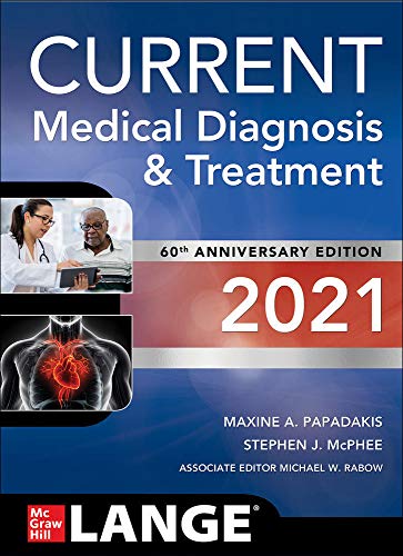
- Publisher:McGraw-Hill Education / Medical; 60th edition (September 10, 2020)
- Language:English
- Paperback:1984 pages
- ISBN-10:1260469867
- ISBN-13:978-1260469868
- ISBN-13:9781260469868
- eText ISBN: 9781260469875
- Item Weight:5.98 pounds
- Dimensions:7.2 x 2.4 x 9.9 inches
ISBN : ISBN : 9781260469868


